clinical_notesClinical Hearing Tests & Audiograms Guide
Everything you need to know about what happens during an NHS or private audiology appointment, how pure-tone testing works, and how to read your personal audiogram.
A professional audiology test is completely painless and usually takes between 30 to 60 minutes. Here are the 5 core steps performed by NHS UHBW audiologists:
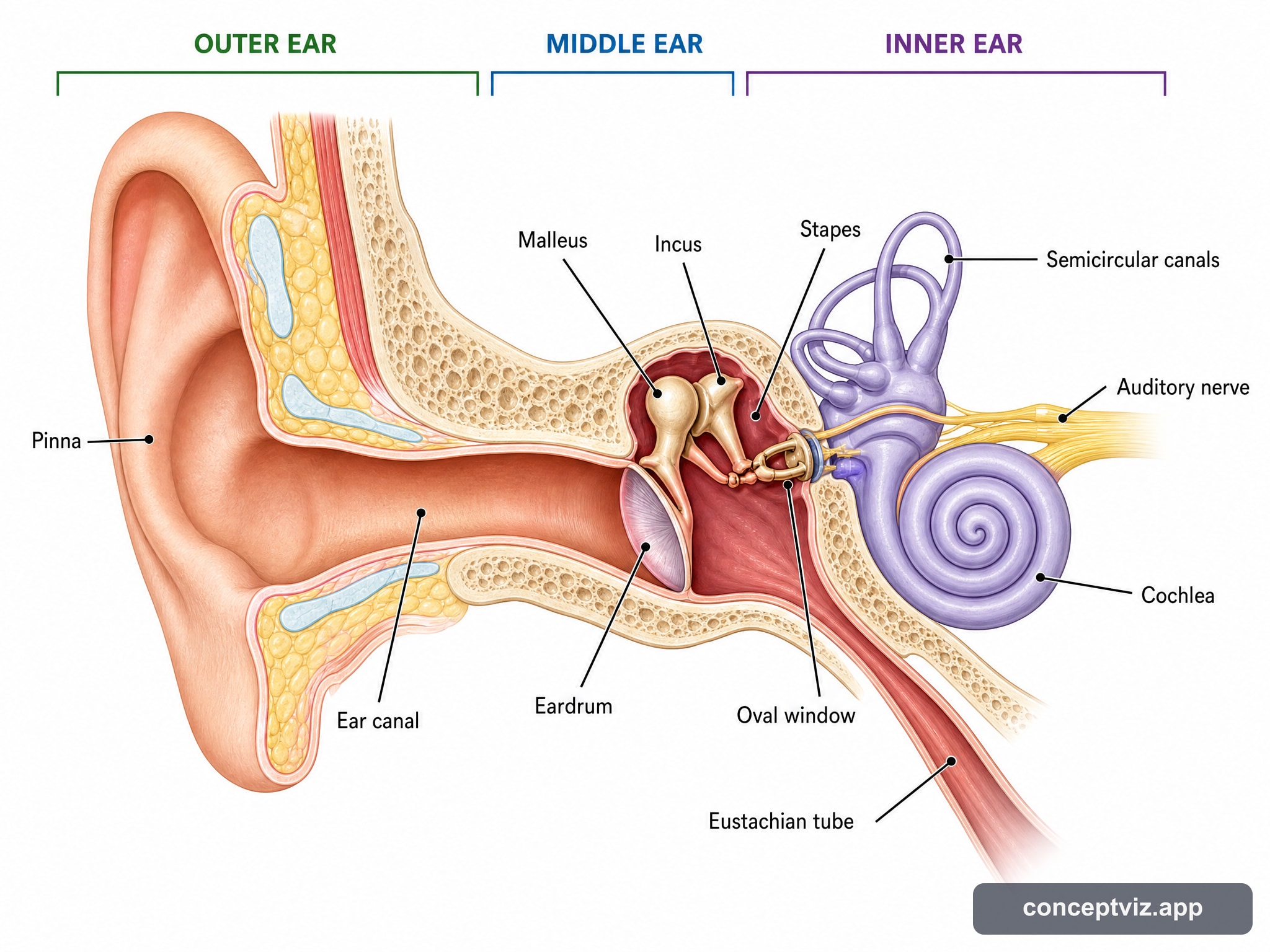
1. Otoscopy Wax Inspection
The audiologist examines inside your ear canal with a lighted otoscope to ensure your ear canals are clear of impacted earwax or infection.
2. Pure-Tone Audiometry (PTA)
Seated in a soundproof booth wearing headphones, you press a button whenever you hear tones ranging from low bass (250 Hz) to high treble (8000 Hz).
3. Bone Conduction Testing
A small vibrating pad is placed behind your ear bone to send sound directly to your inner ear cochlea, distinguishing nerve loss from middle ear blockages.
4. Speech Recognition Testing
You repeat words spoken at comfortable and quiet levels to measure how clearly your brain processes speech in quiet and background noise.
5. Real Ear Measurement (REM) Calibration
The gold standard of hearing aid fitting! A miniature silicone probe microphone is placed in your ear canal alongside your new hearing aid to calibrate output to your ear canal's exact physical shape.
An audiometer is the core diagnostic medical instrument used by audiologists during a hearing test to measure your precise hearing acuity across different frequencies and sound levels.
1. Pure Tone Sound Generator
Produces pure acoustic sine-wave tones at specific pitch frequencies (from low 125 Hz bass to high 8000 Hz treble).
2. Precision Volume Attenuator
Controls sound volume in precise 5 dB increments, from very faint whispers (-10 dB HL) up to loud levels (120 dB HL).
3. Air & Bone Transducers
Routes sounds independently to headphones (air conduction) or a vibrating oscillator on the mastoid bone (bone conduction).
4. Response Button & Plotting
When you press the response switch upon hearing a tone, the audiometer records your quietest threshold and plots it onto your audiogram.
🔍 Clinical vs Screening Audiometers: Clinical audiometers used in NHS hospitals (like UHBW Bristol) include narrow-band masking noise to prevent the non-test ear from "helping" hear tones, ensuring 100% accurate results for each individual ear.
An audiogram is a graph showing the softest sounds you can hear at different frequencies (pitches).
Horizontal Axis: Frequency (Hz)
Runs from left to right like a piano keyboard. Low pitches (250 Hz - deep bass) on the left to high pitches (8000 Hz - whistle/birds) on the right.
Vertical Axis: Loudness (dB HL)
Runs from top to bottom. 0 dB (very quiet whisper) at the top down to 120 dB (painfully loud jet engine) at the bottom. Lower marks mean greater loss.
Universal Audiogram Symbols:
| Hearing Loss Level | Decibel Range (dB HL) | Impact on Speech & Sound |
|---|---|---|
| Normal Hearing | -10 to 20 dB | Hears quiet whispers, clock ticking, and distant speech. |
| Mild Loss | 20 to 40 dB | Struggles with soft whispers and distant background noise. |
| Moderate Loss | 40 to 70 dB | Difficulty understanding conversational speech without hearing aids. |
| Severe Loss | 70 to 90 dB | Requires high-power hearing aids to hear loud voices or traffic. |
| Profound Loss | 90+ dB | Relies on tactile vibration, sign language, or Cochlear Implants. |
In addition to hearing tests, NHS Audiology departments (such as UHBW Bristol Vestibular Clinic) test for inner-ear balance disorders like Benign Paroxysmal Positional Vertigo (BPPV).
What is BPPV?
BPPV occurs when tiny microscopic calcium carbonate crystals (otoconia) break loose from the inner ear balance organ and become trapped inside one of the fluid-filled semicircular canals. Turning your head or rolling over in bed causes the loose crystals to shift, sending false movement signals to your brain and triggering brief episodes of intense spinning dizziness (vertigo).
How Audiologists Test & Treat BPPV:
1. Dix-Hallpike Positional Test
The audiologist guides you from sitting to lying flat with your head turned 45 degrees. If BPPV is present, this movement triggers brief involuntary eye flickering (nystagmus) and dizziness, pinpointing which ear canal is affected.
2. Epley Repositioning Manoeuvre
Treatment can often happen during your appointment! The audiologist takes your head through a sequence of guided physical rotations to coax the loose crystals back out of the canal, resolving the spinning episodes.
- Rest Your Ears: Avoid loud concerts or power tools for 16 hours prior to testing to avoid temporary threshold shift.
- Check for Wax: Ask your GP surgery or local pharmacy to inspect your ears; earwax must be removed before testing.
- List Problem Settings: Write down specific Bristol venues, work meetings, or noisy family gatherings where hearing is hardest.
Know Your Communication Rights at Your GP Practice:
- Double Appointment Time: You can request double-length consultation slots so you don't feel rushed when listening.
- No Surprise Phone Calls: Request that your surgery flags your file for SMS/Email communication only, avoiding missed phone calls.
- Visual Call Screens / Face-to-Face Calling: Request that doctors step into the waiting room rather than calling names over garbled tannoy systems.